
3D Bioprinted Vascularized Kidney Tissue Successfully Transplanted and Functioning in Primate Models

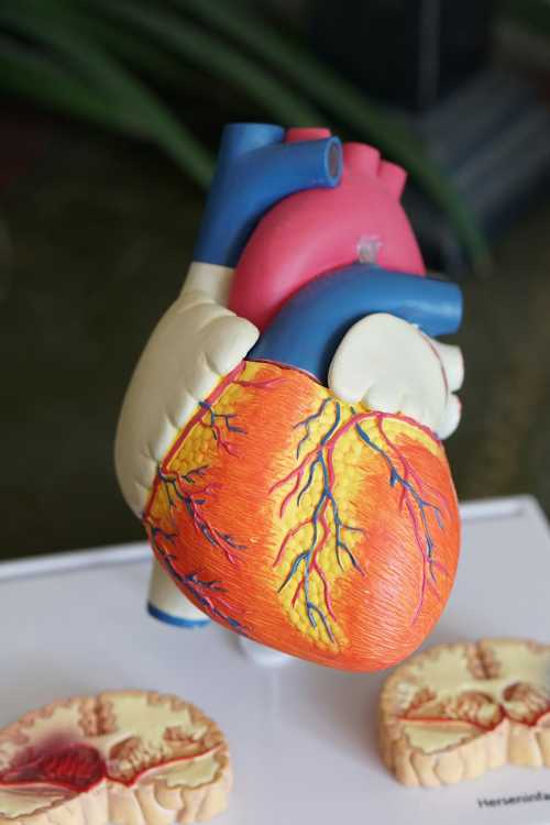
Bridging the Gap Between Organ Shortage and Regenerative Engineering
The critical global shortage of organs for transplantation has been met with a transformative breakthrough as researchers successfully transplanted a fully 3D-bioprinted, vascularized kidney construct into a non-human primate, where it has demonstrated sustained functional filtration for over six months . End-stage renal disease (ESRD) affects millions worldwide, with dialysis serving as a poor long-term substitute for native kidney function and the waitlist for deceased donor kidneys claiming thousands of lives annually. The bioprinted kidney, developed by a consortium of regenerative medicine experts, utilizes a proprietary bioink composed of decellularized extracellular matrix (dECM) harvested from porcine kidneys, seeded with the recipient's own induced pluripotent stem cells (iPSCs) differentiated into renal progenitor cells and endothelial cells . This autologous approach theoretically eliminates the risk of immune rejection, potentially removing the need for lifelong immunosuppressive therapy.
The most significant historical hurdle in bioprinting solid organs has been the creation of a functional, hierarchical vascular network capable of perfusing the thick tissue without causing central necrosis. To overcome this, the research team utilized a novel sacrificial 3D printing technique, printing a highly complex, fractal-like network of carbohydrate glass filaments that were subsequently melted away, leaving behind a perfect, endothelialized microchannel network . When connected to the primate's renal artery and vein, the construct immediately established blood flow. Over the 180-day observation period, the bioprinted tissue demonstrated progressive maturation of the nephron structures. By week 12, the construct was actively filtering blood, producing urine, and clearing creatinine and urea nitrogen, contributing to a 40% recovery of the animal's total renal function after bilateral native nephrectomy.
Histological Maturation and Future Clinical Translation
Histological analysis of the explanted tissue at the six-month mark revealed remarkable structural organization. The bioprinted glomeruli exhibited intact podocyte foot processes and a functional filtration barrier, while the tubular segments showed proper polarity and expression of specific transport proteins necessary for electrolyte reabsorption . The endothelial lining of the vascular channels remained quiescent and non-thrombogenic, expressing appropriate levels of nitric oxide synthase and thrombomodulin. Crucially, there were no signs of fibrotic encapsulation or immune-mediated rejection, confirming the immunological privilege granted by the iPSC-derived autologous cells. The primate maintained normal blood pressure, electrolyte balance, and acid-base homeostasis without the administration of any immunosuppressive drugs.
While the transition from primate models to human clinical trials will require rigorous scaling and long-term safety validation, this achievement represents a watershed moment in regenerative medicine. The ability to manufacture complex, vascularized, and functional organ tissues on-demand using a patient's own cells addresses the two greatest limitations of current transplantation: organ availability and immune rejection. The research team is currently working with the FDA to design the protocols for a first-in-human Phase I safety trial, initially targeting patients with ESRD who are highly sensitized and unlikely to ever receive a compatible donor organ . If successful, 3D bioprinting will transition from a laboratory curiosity to a standard clinical therapy, fundamentally solving the global organ shortage crisis and restoring health to millions of patients awaiting transplants.
Related Articles
The Biopharma Bottleneck: How Supply Chains and Regulatory Reality Are Restraining Medical Innovation
Aug 22, 2026
The Structural Faultlines of Modern Medicine: Navigating the Convergence of Capital, Compute, and Geopolitics
Aug 22, 2026
Comments (0)
No comments yet. Be the first to share your thoughts!
Want to join the discussion?
Please log in to post a comment.
Login NoworCreate an Account